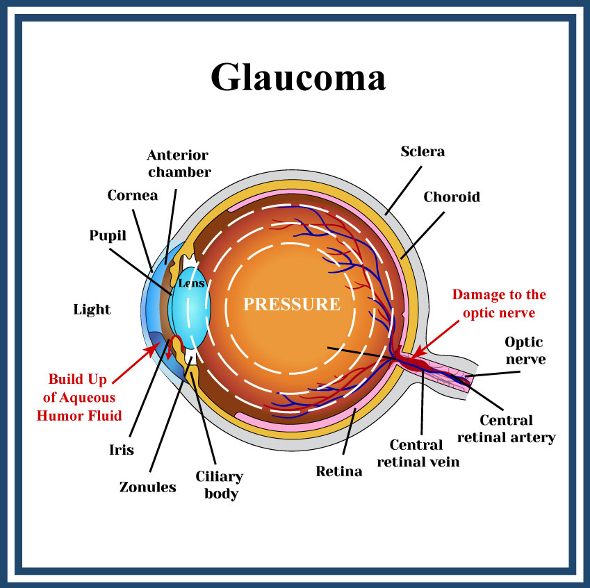
Glaucoma is the leading cause of permanent blindness in the United States, and it is estimated to affect nearly one in every fifty adults. Glaucoma is often called the “silent thief of sight” because in most cases vision loss appears gradually, unnoticed by the patient until it has become severe. Fortunately, with early detection and today’s technology, loss of sight due to most cases of glaucoma can be controlled.

Because most people with glaucoma have no early symptoms or pain, it is important to have regular, routine eye exams so that glaucoma can be diagnosed and treated before long-term visual loss occurs. Early detection and treatment by our doctors at Tampa Eye Clinic is important and is the key to preventing full optic nerve damage and blindness.
Open-angle glaucoma is the most common form of glaucoma, characterized by a gradual increase in intraocular pressure (IOP) due to the slow clogging of the drainage canals in the eye. This condition develops over time and often without noticeable symptoms until significant vision loss occurs.
Narrow-angle glaucoma, also known as angle-closure glaucoma, is a type of glaucoma where the drainage angle formed by the cornea and iris becomes too narrow. This prevents fluid in the eye from draining properly. As a result, intraocular pressure (IOP) increases, which can damage the optic nerve and lead to vision loss.
Laser surgery has become increasingly popular as an intermediate step between medications and traditional glaucoma filtration surgery. Selective Laser Trabeculoplasty (SLT) is a relatively new laser treatment for open-angle glaucoma. SLT uses short pulses of low energy laser light to target melanin-containing cells in a network of tiny channels, called the trabecular meshwork. The objective of the surgery is to help fluids drain out of the eye, reducing intra-ocular pressure that can cause damage to the optic nerve and loss of vision.
The selective technique is much less traumatic to the eye than Argon Laser Trabeculoplasty(ALT), which has been the standard laser procedure. ALT can cause tissue destruction and scarring of healthy cells in the trabecular meshwork structure. SLT reduces intra-ocular pressure without this risk. SLT can be used to effectively treat some patients who could not benefit from ALT. This includes patients who have already been treated with ALT.
Although your doctor may suggest laser surgery at any time, it is often performed after trying to control intra-ocular pressure with medicines. In many cases, you will need to keep taking glaucoma drugs even after laser surgery.
Your treatment will be performed in a specially equipped laser room. It does not require a surgery center.
Once you have been checked in and settled comfortably, drops will be used to numb your eye; no injections or needles are used.
When your eye is completely numb, an eyelid holder will be placed between your eyelids to keep you from blinking.
Your doctor will hold up a special lens to your eye as a high-peak power beam of green light is aimed at the lens and reflected onto the meshwork inside your eye. You may see flashes of bright green or red light. The laser will selectively target melanin-containing cells, resulting in increased fluid outflow. You will not feel any pain during the procedure. It takes 10-20 minutes.
Your eye pressure will be checked shortly after your procedure and drops may be prescribed to alleviate any soreness or swelling inside the eye.
You should relax for the rest of the day.
Follow-up visits are necessary to monitor your eye pressure. While it may take a few weeks to see the full pressure-lowering effect of this procedure, during which time you may have to continue taking your medication, many patients are eventually able to discontinue some of their medications. Most patients resume activities within a few days.
The effect of the surgery may wear off over time. Serious complications with SLT are extremely rare, but like any surgical procedure, it does have some risks. Going to a specialist experienced in SLT can minimize the risks.
If you and your doctor decide that SLT is an option for you, you will be given additional information about the procedure that will allow you to make an informed decision about whether to proceed. Be sure you have all your questions answered to your satisfaction. If you would like more information about this procedure you can make an appointment or contact the office for additional information.
Laser iridotomy is a treatment for narrow-angle glaucoma. In laser iridotomy, a small hole is placed in the iris to create a hole for fluid to drain from the back of the eye to the front of the eye. Without this new channel through the iris, intra-ocular pressure can build rapidly causing damage to the delicate optic nerve, and permanent loss of vision. In most patients, the iridotomy is placed in the upper portion of the iris, under the upper eyelid, where it cannot be seen. The purpose of an iridotomy is to preserve vision, not to improve it.
Your treatment will be performed in a specially equipped laser room. It does not require a surgery center. Once you have been checked in and settled comfortably, drops will be used to numb your eye; no injections or needles are used.
First, your ophthalmologist will place a drop in your eye to make your pupil smaller. This stretches and thins your iris, which makes it easier for the laser to make the pinhole sized puncture.
Next your doctor will place a special contact lens on your eye to focus the laser light upon the iris. This lens keeps your eyelids separated so you won’t blink during treatment. It also reduces small eye movements so that you don’t have to worry about your eye moving during the treatment.
To ensure that the contact lens doesn’t scratch your eye, a special jelly will be placed on the surface of your eye. This jelly may remain on your eye for about 30 minutes, leading to blurred vision or a feeling of heaviness.
During the laser treatment, you may see a bright light, like a photographer’s flash from a close distance. Also, you may feel a pinch-like sensation. Other than that, the treatment should be painless.
Your eye pressure will be checked shortly after your procedure and drops may be prescribed to alleviate any soreness or swelling inside your eye.
Follow-up visits are necessary to monitor your eye pressure. Your doctor may ask you to continue using eye drops to make your pupil smaller for a few days following your laser treatment. These drops can temporarily cause blurred vision (especially at night) and may also give you a slight headache. Your doctor may use other drops, both before and after your treatment to control your eye pressure. Still other eye drops may be used to reduce inflammation.
Everyone heals differently, but most people resume normal activities immediately following treatment, although you’ll need to have someone drive you home after your procedure. For the next few days your eyes may be red, a little scratchy and sensitive to light.
Serious complications with laser iridotomy are extremely rare, but like any medical procedure, it does have some risks. The chance of losing vision following a laser procedure is extremely small. The main risks of a laser iridotomy are that your iris might be difficult to penetrate, requiring more than one treatment session. Another risk is that the hole in your iris will close. This happens in less than one-third of the cases.
Following your procedure, you may still require medications or other treatments to keep your eye pressure sufficiently low. This additional treatment will be necessary if there was damage to the trabecular meshwork prior to the iridotomy or if you also have another type of glaucoma in addition to the closed-angle type.
If you would like more information about this procedure you can make an appointment or contact the office for additional information.
Filtration surgery, also called trabeculectomy, is a treatment for several types of glaucoma including open-angle and narrow-angle glaucoma. It is often performed on patients who have not responded well to medication or laser treatment such as ALT or SLT. Filtration surgery usually provides a dramatic reduction in pressure within the eye. A small channel, or ‘bleb’ is created to allow fluid to drain from the eye.
You will arrive at the surgery center 30-60 minutes prior to your procedure. Once you have been checked-in and settled comfortably, you will be prepared for surgery.
The area around your eyes will be cleaned and a sterile drape will be applied. You may be given a sedative to help you relax.
Your eye will be numbed with topical or a local anesthesia. When your eye is completely numb, an eyelid holder will be placed between your eyelids to keep you from blinking.
Using advanced microsurgical techniques and equipment, your doctor will create a tiny new channel between the inside of your eye and the outside of your eye.
A small section of tissue will be removed, creating a channel, to allow fluid to pass through the blocked drainage network onto the white (sclera) of the eye.
The incision will be closed with small stitches and covered with the thin outer tissue of the eye, called the conjunctiva. Blood vessels in the conjunctiva will carry the draining fluid away.
To keep the drainage channel open, your doctor may apply an extremely small dose of a chemotherapeutic agent to the new filter.
Your eye pressure will be checked shortly after your procedure and drops may be prescribed to alleviate any soreness or swelling inside the eye.
You should go home and relax for the rest of the day. Most patients resume normal activities within a few days.
Follow-up visits are necessary to monitor your eye pressure. It may take a few weeks to see the full pressure-lowering effect of this procedure, and adjustments may need to be made to the filter during this period. These adjustments may include:
The success rate for this type of surgery is approximately 80 percent in cases where no surgery has been done on the eye before. However, everyone’s eyes are unique and many people do require further treatments. In more difficult cases where even filtration surgery doesn’t prevent damage to the ocular nerve, it may be necessary to perform other types of procedures.
Serious complications with filtration surgery are extremely rare, but like any surgical procedure, it does have some risks. Going to a specialist experienced in filtration surgery can significantly minimize the risks.
If you would like more information about this procedure you can make an appointment or contact the office for additional information.
Because most people with glaucoma have no early symptoms or pain, it is important to have regular, routine eye exams so that glaucoma can be diagnosed and treated before long-term visual loss occurs. Early detection and treatment by our doctors at Tampa Eye Clinic is important and is the key to preventing full optic nerve damage and blindness.
Glaucoma occurs when pressure builds in the eye. A liquid called aqueous humor constantly flows through the eye and through a drainage system. When the drainage system becomes blocked, aqueous humor builds up and puts pressure on the optic nerve. If too much pressure builds on the optic nerve, vision loss can occur.
